Hello everyone, how are you all? Hope you all are doing well and its been long about 2 months i think i didn't made a post and didn't get interacted with you all because of my busy schedule.
But i got some little break now. I would like to tell about one pathological finding that we will see whenever we get exposure to many cases. The finding name is called "Monckeberg medial calcific sclerosis".
Where we will see it?
The answer for this question is in the tunica media of small to medium sized blood vessels.
What is calcification?
Its in simple words is calcium deoosition
The type of calcification is dystrophic calcification.
There are mainly two types of calcification one is Dystrophic calcification and another one metastatic calcification.
Dystrophic calcification
In this, the deposition of calcium occurs in degenerated or dead tissues. Monckeberg medial calcific sckerosis comes under in this category. The calcium levels will ve normal.
Metastatic calcification
In this, the deposition of calcium occurs in normal tissue. The calcium levels are deranged and hypercalcemia is seen. Example is hyperparathyroidism.
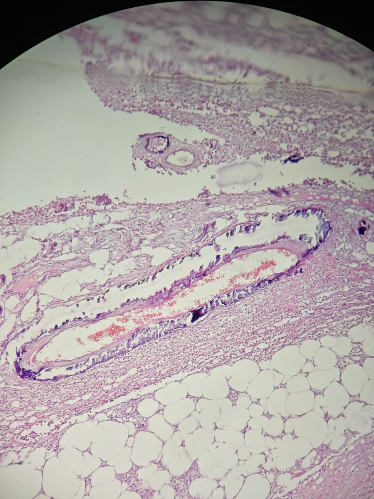
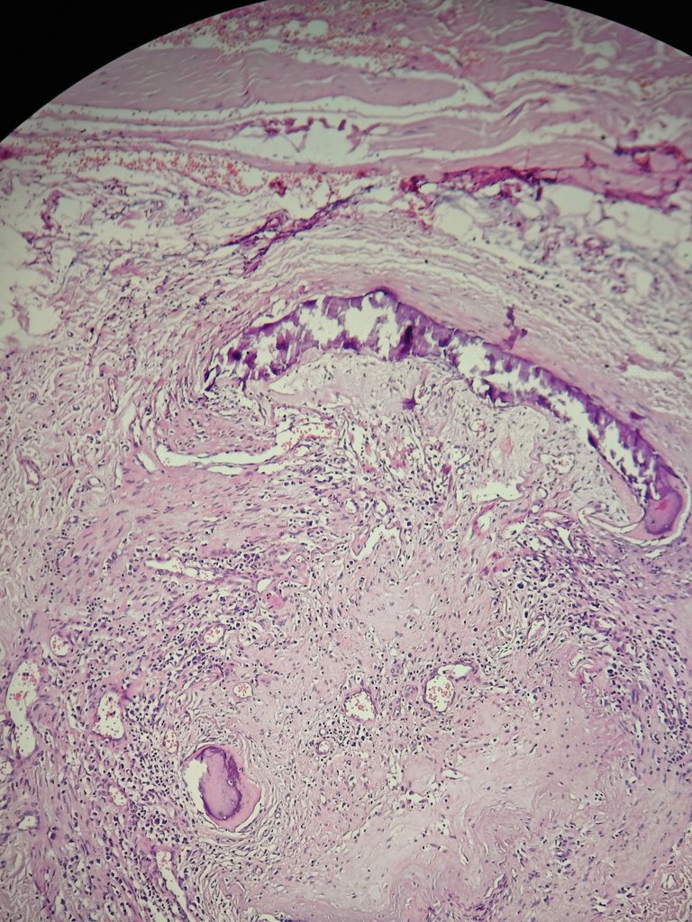
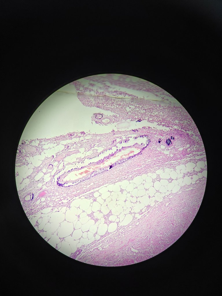
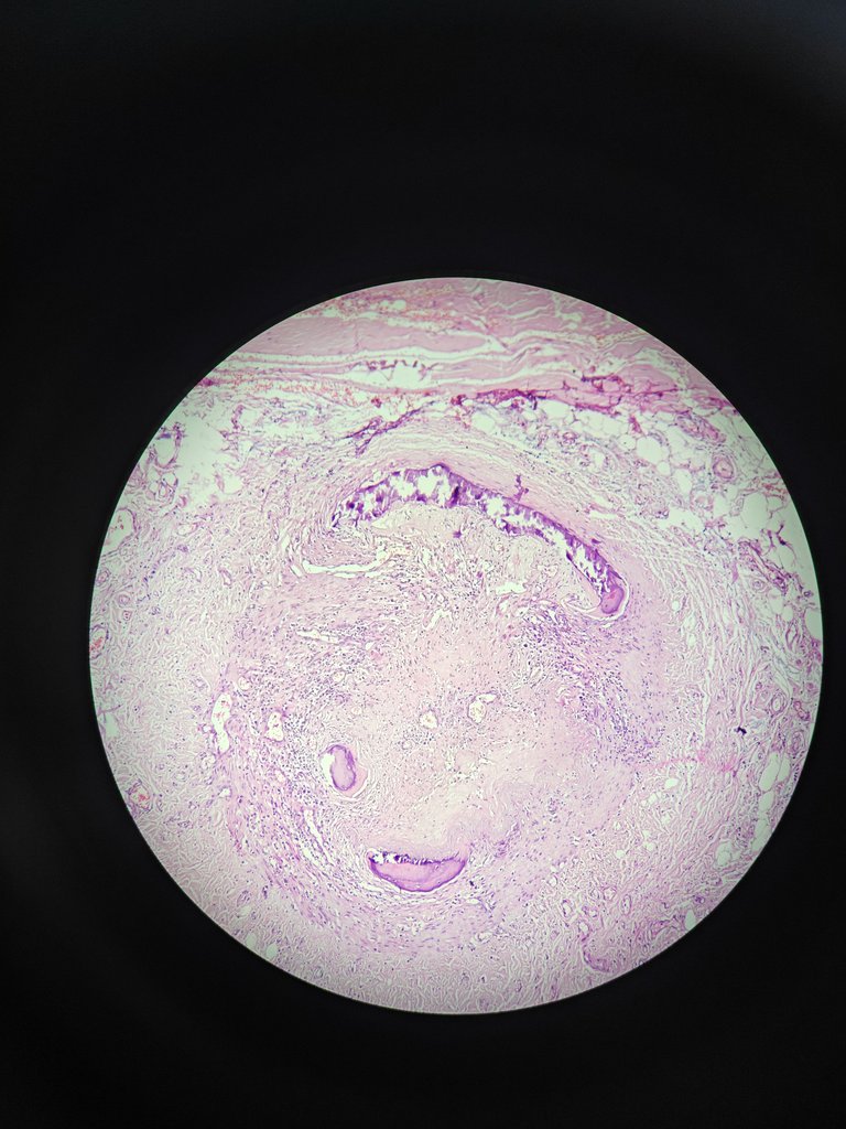
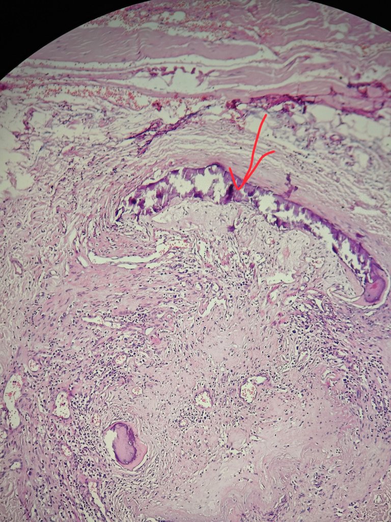
Histopathological images
You can in the below image.
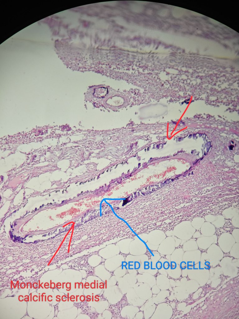
The RBCs represent the vessel lumen where they are present. The surrounding is vessel. The wall of blood vessel has bluish color deposition which represents calcium.
This is called monckeberg medial calcific sclerosis.
References
Rosai and Ackerman's Surgical Pathology - 11th Edition
Hope you have learned something today.
Thanks for reading,
With regards,