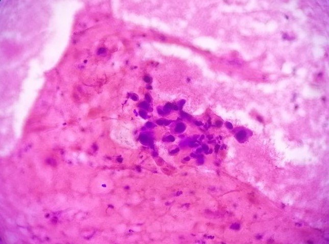
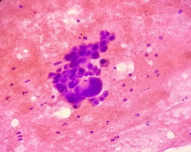
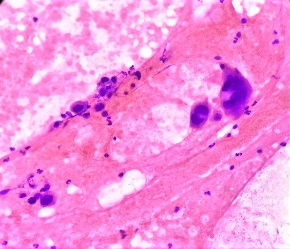
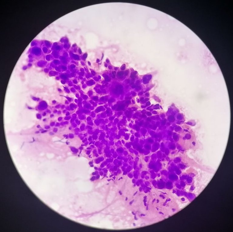
A case of an 81 year old male previously diagnosed as having Squamous Cell Carcinoma (SCC). The specimen below is a fine needle aspiration for a left axillary mass suspected to be a tumor recurrence. Even if an excision biopsy was done to remove the tumor locally, the chance that metastasis has already occurred can't be ruled out.
Appreciate what malignant cells look like~
Taken at 400x magnification under H and E stains:


Taken at low power magnification 100x:

Hyperchromatic, round to markedly pleomorphic nuclei with inconspicuous nucleoli and scant to abundant eosinophilic cytoplasm.
Features that makes those cells look really malignant are described above, but another simple way to put it is the dark nucleus being "hyperchromatic" means it's really active and ready to divide, attaching the word pleomorphic means it's assuming an irregular to bizarre forms. Some giant forms really make this case easy to call.
I just said it has an inconspicuous nucleoli because I couldn't appreciate the nucleoli on the stains but generally, if a cell is reactive or just kicking its cellular machinery into high gear, you'll see prominent nucleoli within the nucleus. This is readily seen on tissue sections. The scant to abundant cytoplasm just adds weight to the increase in nucleus to cytoplasmic ratio commonly expected from malignant cells.
The red background is made up of mature blood elements (RBCs, and mixed inflammatory cells, necrotic debris, fibrin and hemorrhage.) SCC is often has an abundant necrotic debris. I was also looking for keratin flakes and pearls on the background to further raise my case as this being a Squamous Cell Carcinoma. The ideal way to diagnosed these type of cases was examining a tissue section so calling it definitively as SCC requires a lot of guts given how it's just probably a small sample overall. There's still a chance that it could be Adenosquamous Cell Carcinoma (a different disease entity but that's beyond the scope of this blog).
Seeing these clusters of cells made me instantly call it malignant and favoring SCC recurrence. It's not often do I get to see a clear cut picture that some set of cells are malignant especially if they came from well differentiated tumors. Well differentiated gives me an idea they look close to the normal ones in operation.
Based from these findings, I'd say it's consistent with a moderate to poorly differentiated squamous cell carcinoma. The patient was previously diagnosed as moderately differentiated SCC. Giving more weight that this is a recurrence and not some other disease process at work.
For cases like these, the likelihood of the patient just undergoing palliative care is high given how the age of the patient and health status may pose little benefit from chemotherapy. Recurrence also raises the possibility that this set of neoplastic cells are resistant to the the previous chemo drugs used (if the patient did undergo chemotherapy). Their body may not be able to handle the chemo drugs.
There are two sides of looking at this case from my point of view:
- I get excited to see first hand what the textbooks say. I'm applying what I learned and this is an even higher form of education.
- At the end of the day, someone is suffering for this learning experience.
This is how I looked at all the interesting cases I've encountered.
I own the photos. You stumbled upon my medical blog.
If you made it this far reading, thank you for your time.
Posted with STEMGeeks


