The specimen came from a 46 year old female that complained about a bleeding polyp. When I received the specimen, it just looked like any other tissue that had a dark brown to black appearance which I just attributed to hemorrhage. This is the second time I encountered such case.
The lens used for taking the 400x needed some cleaning and we had to wait for the yearly maintenance to come in when I took the shots. I had adjusted the photos to get the details as close as possible.
Take at 40x magnification:
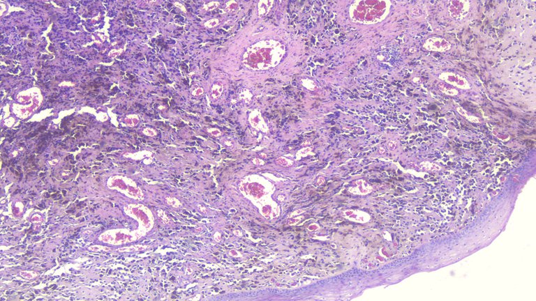
There's the overlying skin and underneath are sheets and cords of malignant cells formed.
Take at 100x magnification:
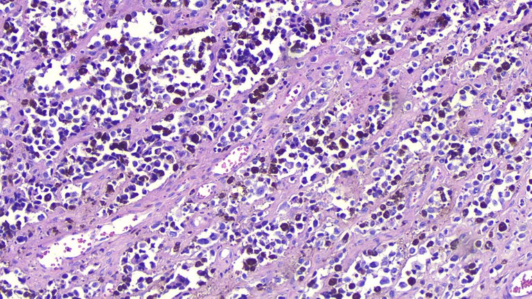
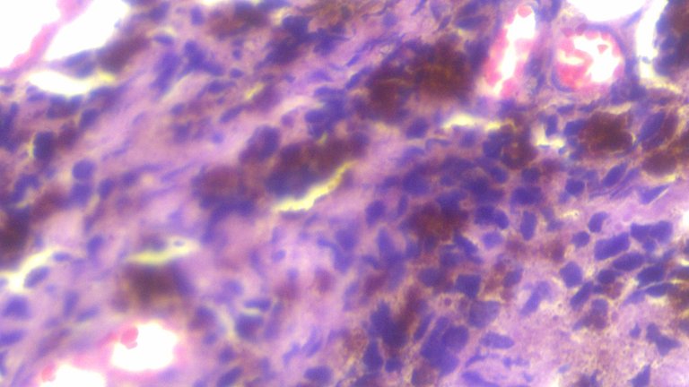
Taken at 400x
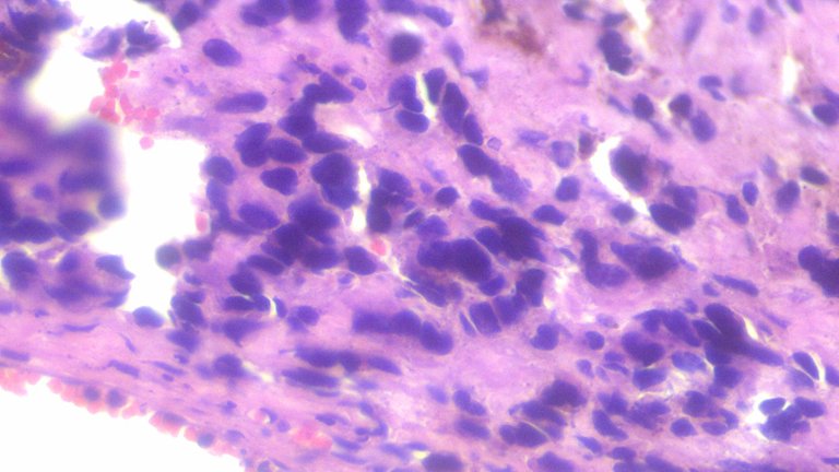
Some malignant cells don't display pigment but you can still see some scant pigments on this shot. There are cases when you got nothing like that on the slide and you're left with these hyperchromatic cells with irregularly shaped nuclei to work with. This gives you a bucket list of possibilities to narrow down.
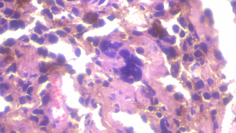
This is a bizarre nuclei way different from the usual round to ovoid neoplastic cells you see surrounding.
I shared another case of melanoma coming from the thigh and the pictures have a lot of similarities.
Some things to keep in mind when seeing pigments are the distribution, color and the cells producing them. You would see yellow to brown pigments when looking at a normal liver tissue because that's where bile is supposed to be made. You would see brown pigments on hemosiderin-laden macrophages because macrophages take up iron for recycling where there is hemorrhage.
Pigments seen when there aren't supposed to be is a good tell. Now seeing cases of melanoma exhibiting these pigments helps narrow down some differentials but there are cases where melanoma doesn't show any pigments and just look like any other poorly differentiated neoplasm. That's where immunohistochemical stains become helpful in eliminating or ruling in other diagnosis.
The range of how melanoma presents itself has dubbed it as one of the great mimickers because it may present itself morphologically as something else when you're least expecting it to be.
We still recommend doing immunohistochemical stains (HMB-45 and S100) as a confirmatory test but base don morphology alone, it most likely is what it is.
Now discussing treatment and prognosis is out of the scope of this post. Melanomas are malignant and highly aggressive. These are also known to do skip metastasis where you can have it on your hand and it also spread to the nose (in contrast to most tumors that spread locally).
If you made it this far reading, thank you for your time.
Posted with STEMGeeks
