Hello everyone, how are you all? Its been so many days i was faraway from @stemsocial and due to my busy schedule and work i don't have time to share. But i decided to share the clinical cases and how diagnosis has been made on it. Let me tell about myself about what i am doing. I am pursuing my MD course in Pathology in AIIMS Mangalagiri and now i am in 1st year and i would like share every case.
Case
This is a case of 31 years female patient came with complaints of small lump in the left breast since 2 year which gradually increased to present size.
On examination - lump size is 4x3 cm, firm in consistency, mobile and associated with pain.
Then i did FNAC(Fine Needle Aspiration Cytology) on the lump present in the left breast. I did 1 pass. Then got material into syringe. Nature of aspirate is blood mixed. Then we spread the aspirated material on the slides and we got 3 slides. Then staining was done. 3 stains were done. Giemsa, H&E and pap.
After staining i have seen under microscope and this is how i got.
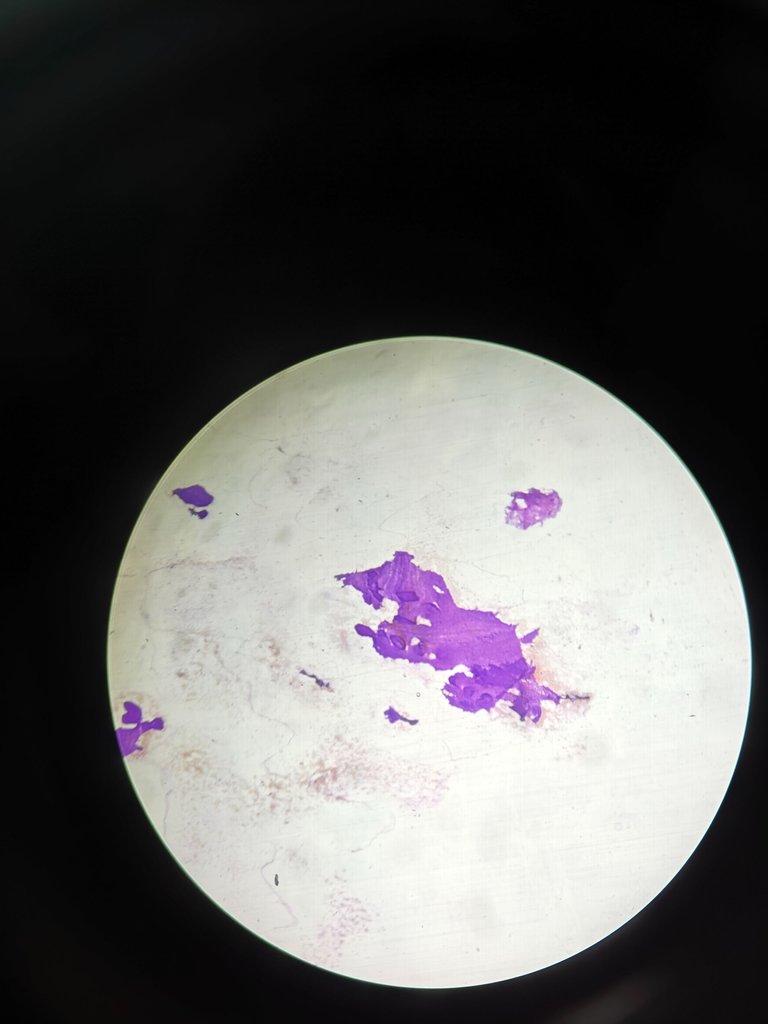
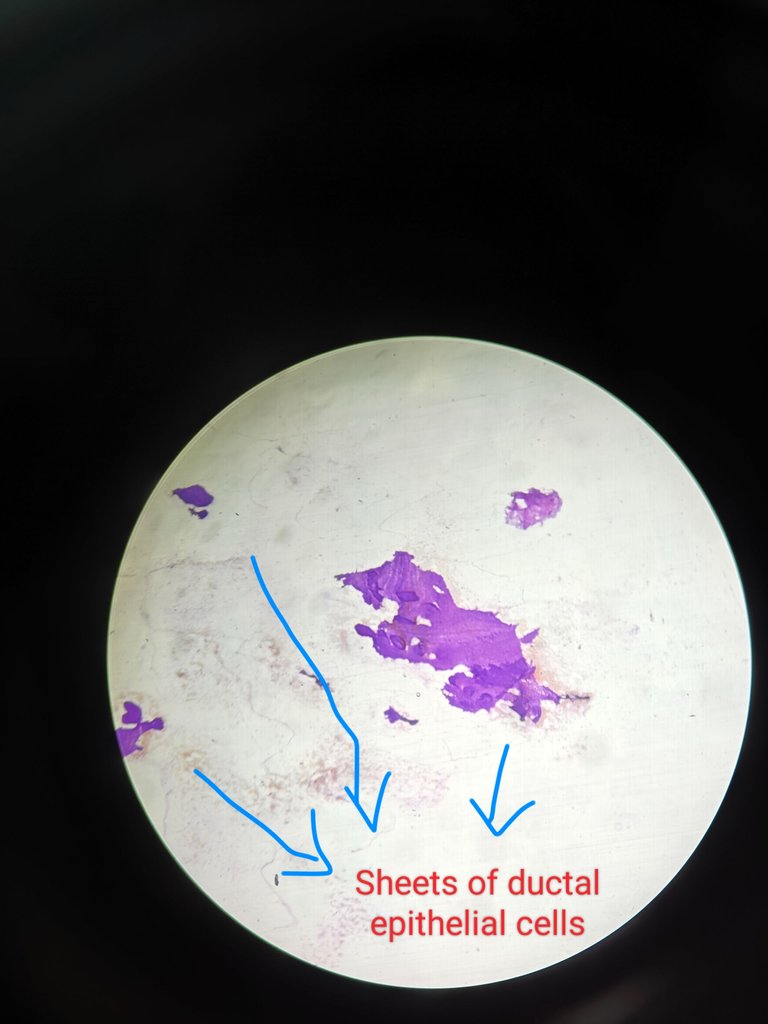
Microscopic findings
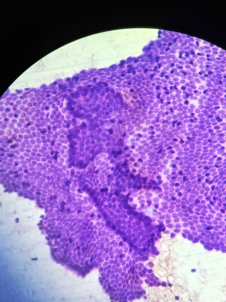
4X
On 4X magnification we can see that there are sheets of cells. These are benign ductak epithelial cells. They form the cohesive monolayered sheets. Some are in staghorn or antler horn pattern also. You can see in the images that i shared.
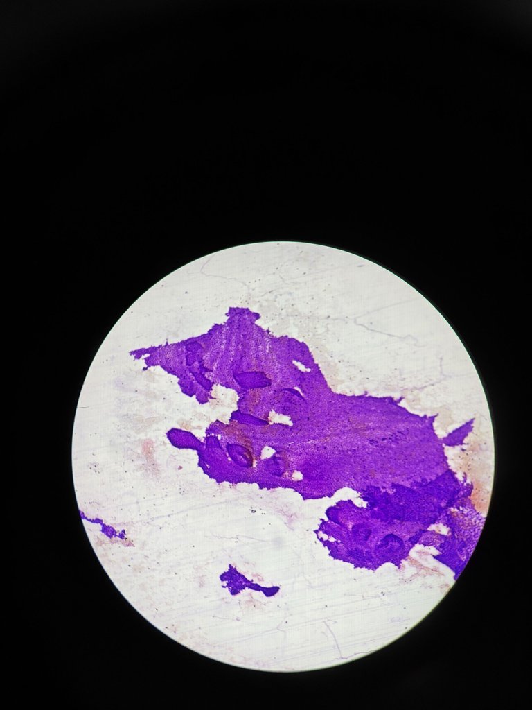
10X
On 10X magnification you can see the cohesive monolayered sheets. Antler horn pattern/staghorn pattern also you can see.

20X
On 20x magnification you can see monolayered sheets and small monolayered sheets on top of it.
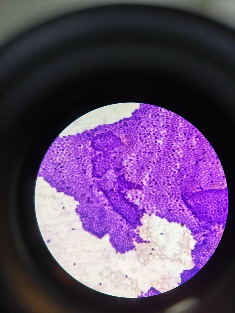
40X
On 40x magnification you can see there are dark blue color cells. Which are singly scattered they are present on top of the monolayered sheets and these are called myoepithelial cells. There is myoepithelial rimming also seen which are surrounding the monolayered sheets of ductal epithelial cells.
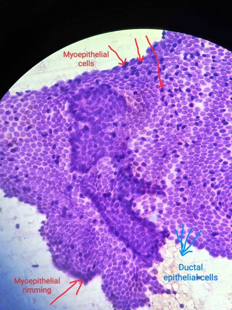
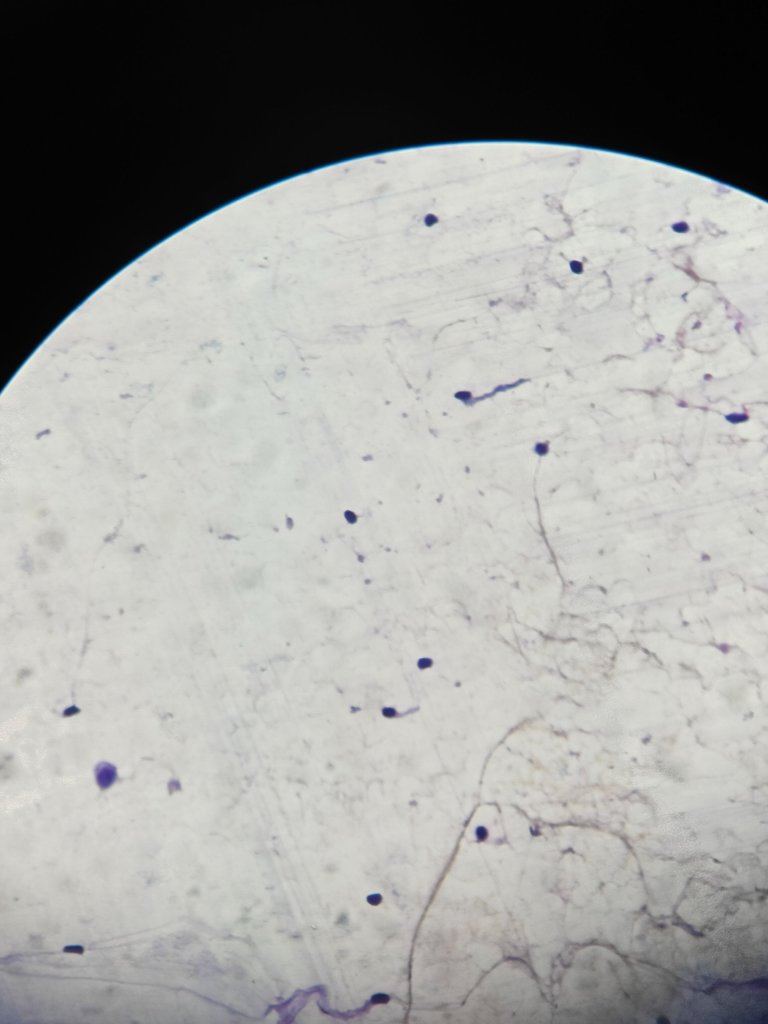
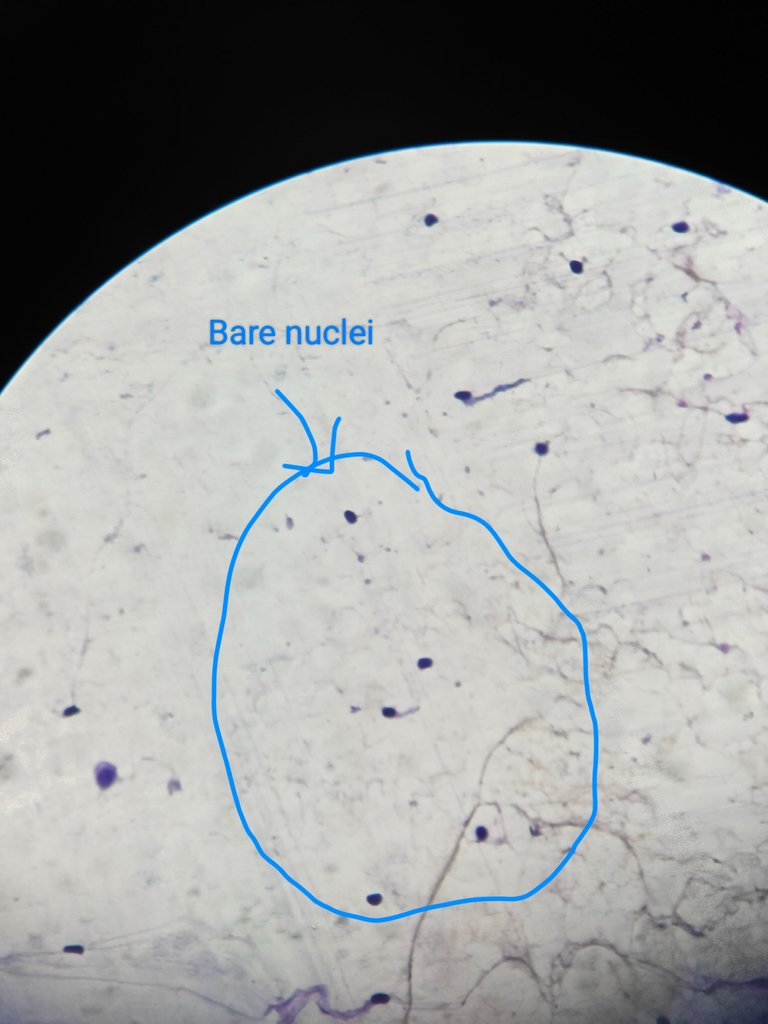
In the back ground there singly scattered bare nuclei which are seen and these are myoepithelial cells. Some times there are bipolar nuclei also seen. Few stromal fragments also seen along with it but here i didn't get stromal fragments because i did only one pass of fnac.
If you see and find these things on microscope then you can give report as FIBROADENOMA.
Its comes under category II(Benign)
(The International Academy of Cytology (IAC) Yokohama System for Reporting Breast Fine-Needle Aspiration Biopsy (FNAB) Cytology)
Some people may say Benign breast disease which is a broad term used where fibroadenoma also comes in it if they don't find stromal fragments.
Fibroadenoma is a benign disease it occurs most commonly in young age females(18-25 yrs)
This is how fibroadenoma diagnosis is made on FNAC.
Thanks for reading,
Yours summisimeon
References -
Robbins & Cotran Pathologic Basis of Disease textbook 10th edition
The International Academy of Cytology Yokohama System for Reporting Breast Fine Needle Aspiration Biopsy Cytopathology