Hello everyone, how are you all? Hope you are doing well. I am too. As i was very bust with residency and i am not getting enough sleep( just sleeping 4 or 5 hrs max), i am not regularly making posts. But from now onwards, i would like to be regular on hive. Today I come up with an interesting thing. I learnt this during my residency and i want to share with you all.
This is all about fluids. In our body, there are many fluids, for example you can take pericardial fluid, pleural fluid, ascitic fluid, cerebrospinal fluid etc.... pulmonary medicine is the department which deals with lung diseases. So they will send pleural fluid most commonly. The question is why they will send?? Lets take an example one patient came to hospital with history of cough,breathless etc.... the pulmonary doctor will think of some differentials like it could be pulmonary TB, fungal infection, malignancy etc.... our main concern here is malignancy which is cancer.
So suppose if there is cancer in lungs it will spread into pleural fluid. So if we examine pleural fluid we can tell the patient has lung cancer or not.
case
So here one patient of 50 yrs age with history of smoking for 20+ years came to hospital with complaints of cough, breathlessness and weightloss and pulmonary doctor examined the patient and xray was done and finally came to know that pleural fluid was increased in volume and they have taken pleural fluid of 50 ml and sent it to pathology department for Fluid cytology to know whether its infection related or malignancy. So we centrifuged fluid and we made smears from the sediment. Then i examined the smears under the microscope. Then these are the things i have seen.
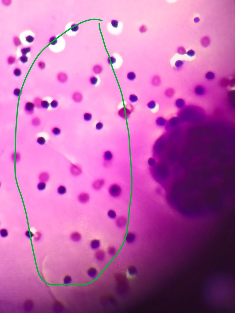
The above image with green color labelling represents lymphocytes and hemorrhage in the background of smear.
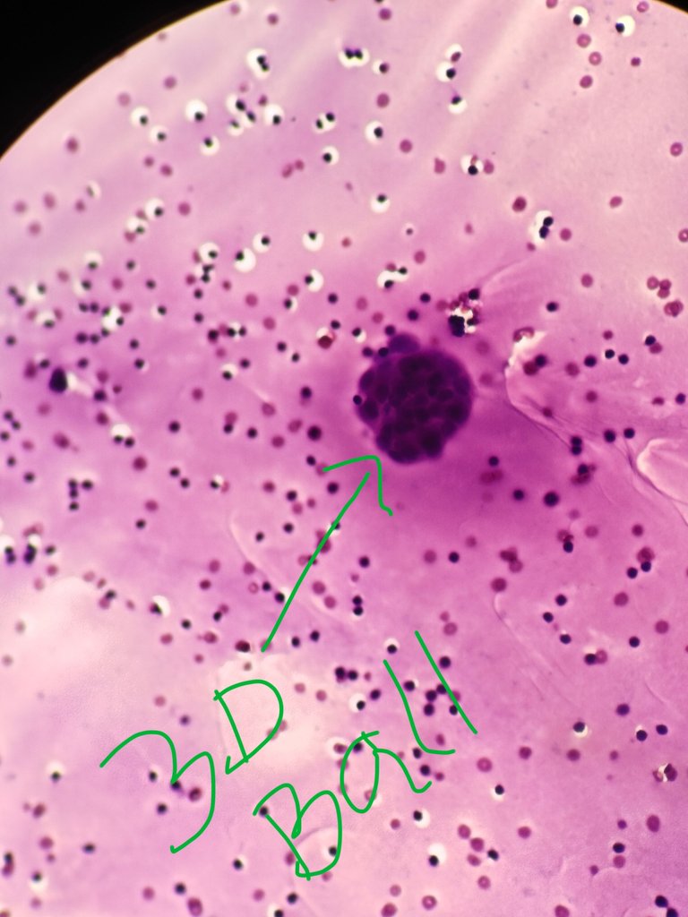
The above image the green labelled cluster is the cluster of tumor cells or atypical cells. Normally we won't see this type of 3D ball like clusters in fluid cytology. If we are seeing like this means there is strong suspicion of malignancy. We should be careful with mesothelial clusters too because they also makes these clusters. But, tumor clusters have atypical nuclear features. Thats why careful observation is much needed.
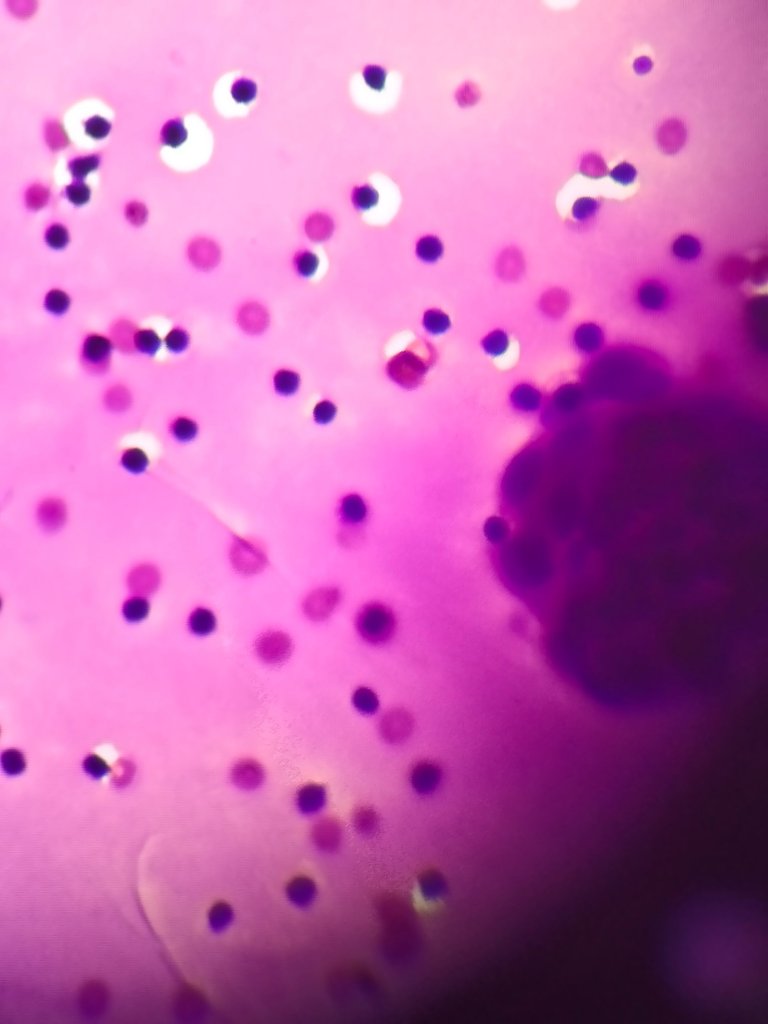
You can see below images in different magnifications there are 3D ball like clusters. So this is a clue that it is positive for malignancy.
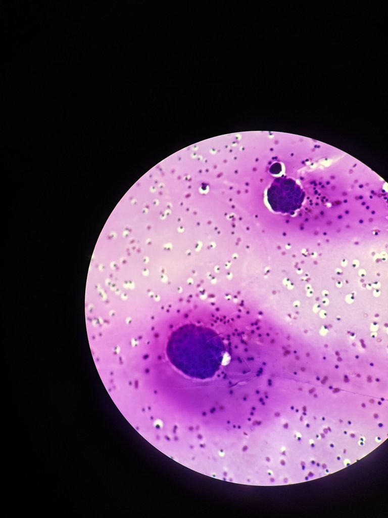
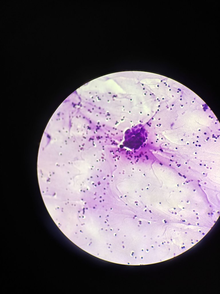
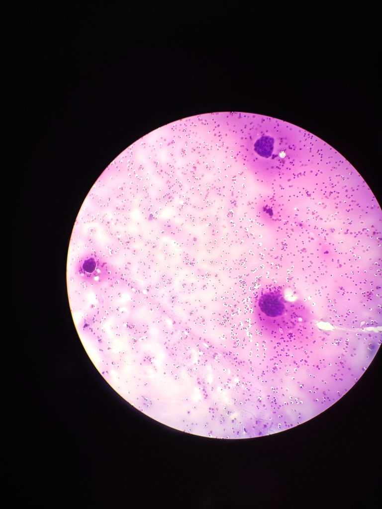
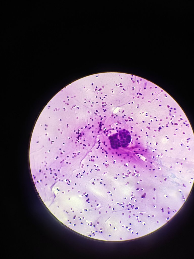
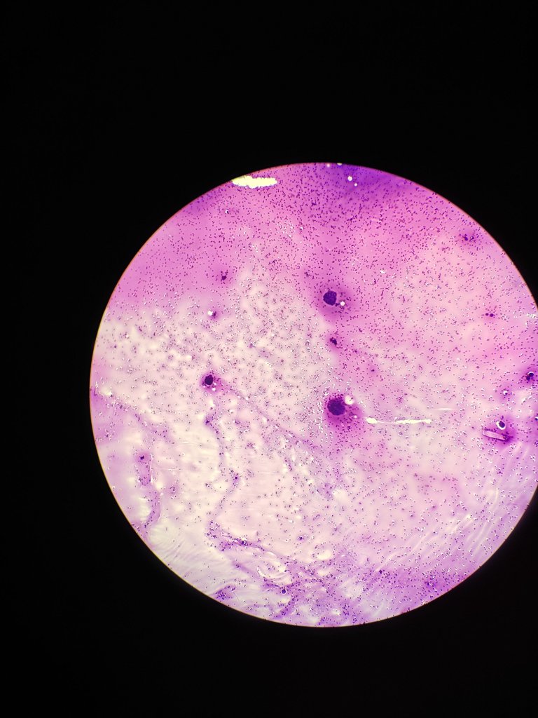
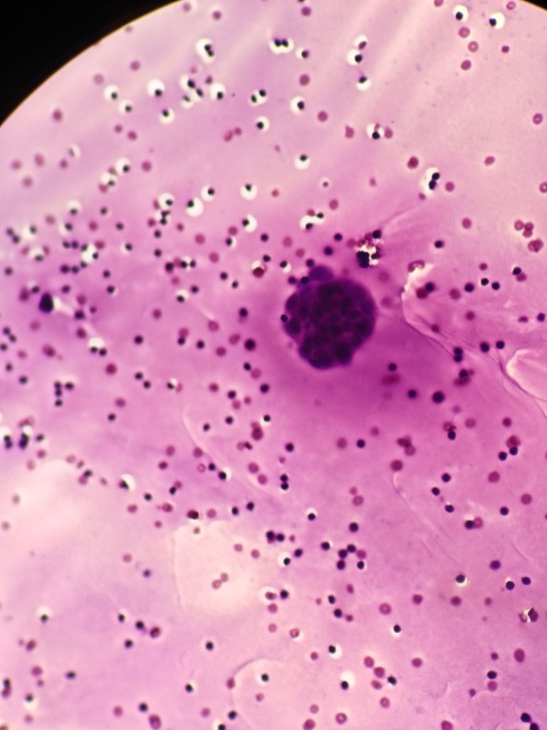
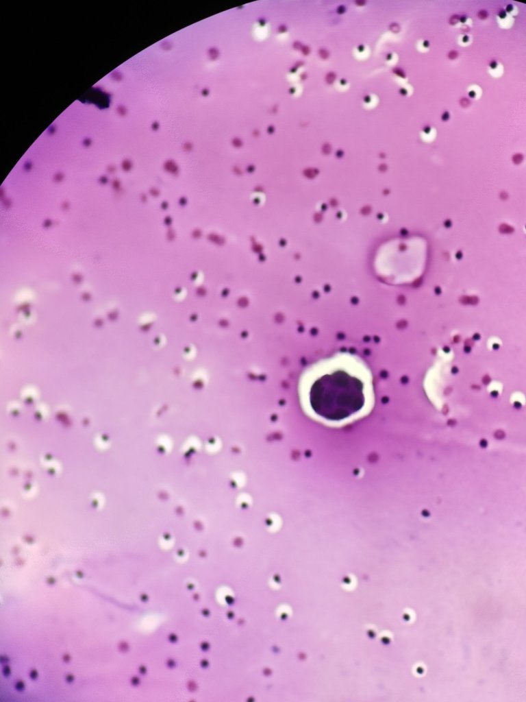
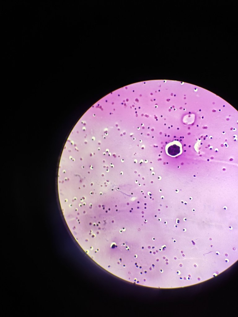
Adenocarcinoma we can consider if we see these findings in smears. But cell block and IHC is confirmatory. So we can suspect primary tumor is from lungs by clinical radiological correlation.
So fluid cytology helps in diagnosing malignancy and patient can be early diagnosed and treatment can be started after pathological, radiological and clinically correlated.
So if you find any of your family member with pleural effusion, you can send pleural fluid to look for malignancy. Not only pleural fluid but other fluids too like Asicutic fluid for ascitis.
Thanks for reading,
With regards,
References
- The International System for Serous Fluid Cytopathology
- Atlas of Serous Fluid Cytopathology
A Guide to the Cells of Pleural, Pericardial, Peritoneal and Hydrocele Fluids


