Hello everyone,how are you all? Hope you are doing well.we learnt about abdoment and its regions and inguinal canal in last two posts. Now as continuation of it we are going to learn about rectus sheath.
RECTUS SHEATH
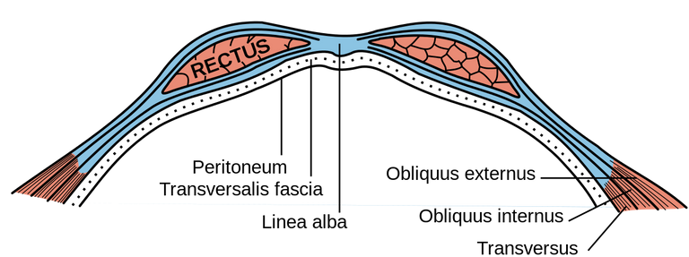
The rectus sheath (also called the rectus fascia) is a tough fibrous compartment formed by the aponeuroses of the transverse abdominal muscle, and the internal and external oblique muscles.
The rectus sheath contains -
1)2 muscles - rectus abdominis muscle and pyramidalis muscle
2)2 vessels - superior epigastric vessels and inferior epigastric vessels
3)lower 6 thoracic nerves(T7-T12)
T7-T11 are intercostal nerves and T12 is called subcostal nerve*
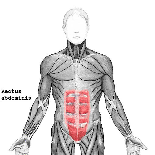
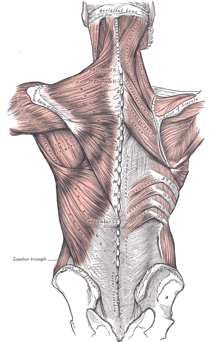
The six packs we get or we see is the rectus abdominis muscle . In the below image you can see it.

Now we have to know about arcuate line.
Arcuate line is present miday between umbilicus and pubic symphysis on either side.you can see in the below image which is labelled as linea semicicularis
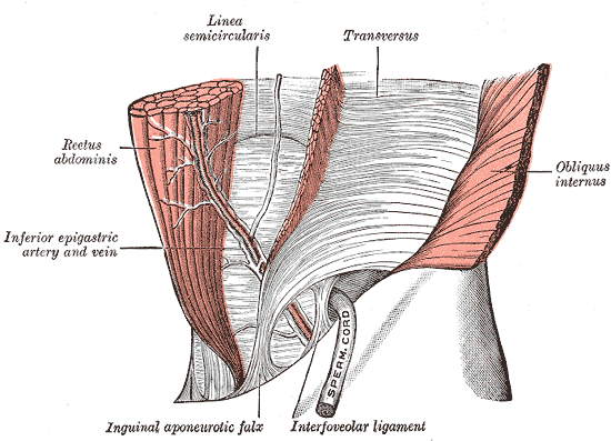
Why should we have to learn about it? Because above the arcuate line & below the arcuate line, the aponeurosis of these muscles are arranged differently.
Above the arcuate line
Each aponeurosis has 2 laminas- anterior lamina and posterior lamina.
Above the arcuate line anterior lamina of internal oblique aponeurosis is present anterior to rectus abdominis muscle and posterior lamina is present posterior to rectus abdominis muscle.
So above arcuate line anterior wall is formed by external oblique aponeurosis and anterior lamina of internal oblique aponeurosis.posterior wall is formed by posterior lamina of internal oblique aponeurosis and transversus abdominis aponeurosis.
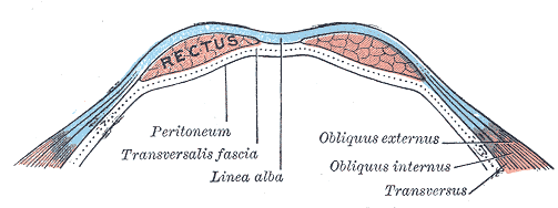
Below the arcuate line
Here the arrangement is different.anterior wall is formed by aponeurosis of external oblique,internal oblique and transversus abdominis.you can see in the below image
Thoracolumbar fascia
The thoracolumbar fascia
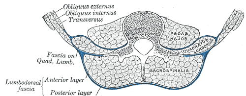
(lumbodorsal fascia or thoracodorsal fascia) is a complex,multilayer arrangement of fascial and aponeurotic layers forming a separation between the paraspinal muscles on one hand, and the muscles of the posterior abdominal wall (quadratus lumborum, and psoas major) on the other.
You Can see in the below image the blue color one which separates psoas major and quadratus lumborum muscles which are abdominal muscles present in the posterior abdominal wall and paraspinal muscles(muscles which are present one either side of spine) which is erector spinae muscle or sacrospinalis muscle.
REFERENCES
Gray's Anatomy for students,3e page no 286,287
Gray's Anatomy:The Anatomical Basis of Clinical Practice,41st Edition Fig 61,page no 1070,1075
By this post, we learnt about half of anatomy of abdomen and i tried to explain with images i request you to please go through my previous posts so that you can understand them in sequence.
Thanks for reading,
With regards,
