Hello everyone, how are you all? Its been a long time that i am not in hive due to my exams and now i finished amy exams and i want to restart & continue again my medical blogs again.
I am going to start on anatomy of abdomen.learning about abdomen anatomy is so important why because many important visceral organs and blood vessels are present in it. Ok lets enter into abdomen.
ABDOMEN
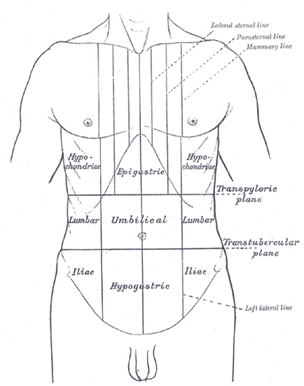
Abdomen is divided into 9 regions by 4 planes.these planes are 2 vertical planes and 2 horizontal planes.
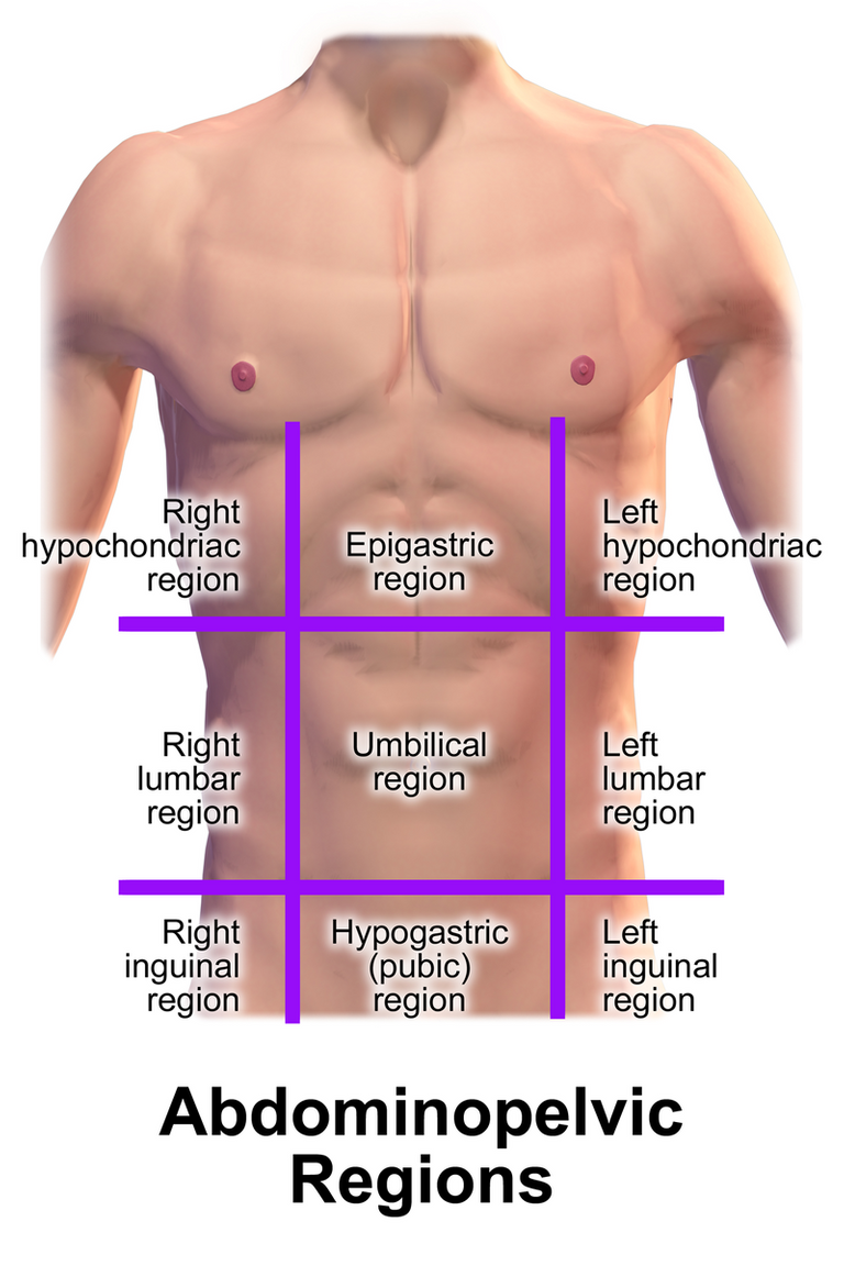
The 2 vertical planes are imaginary lines passing downwards in midclavicular plane.
The 2 horizontal planes are - subcostal plane and transtubercular plane.
Subcostal plane passes through lower border of 2nd Lumbar vertebrae and transtubercular plane passes through upper border of 5th Lumbar vertebrae.
You will understand everything in the above diagram.
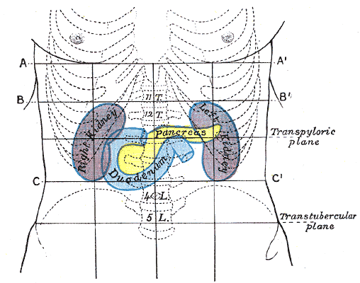
There are two more horizontal planes passing are transpyloric plane which passes through lower border of 1st lumbar vertebrae and supracristal plane which passes through body of 4th lumbar vertebrae.
Why should we have to learn about transpyloric plane is because many important structures lie in this plane.lets see what are the structures that present in this plane-
- Pylorus of stomach
- Spinal cord termination
- Origin of superior mesenteric artery
- Hilum of kidney
- Fundus of gall bladder
- Tip of 9th costal cartilage
Anterior abdominal wall
Abdomen has 8 layers that acts as wall and protect visceral organs which are present inside the abdomen.
The 8 layers from outside to inside are -
- Skin
- Superficial fascia it has two layers outer fatty layer called - "CAMPERS FASCIA" and inner fibrous layer called "SCARPA FASCIA"
- External oblique muscle
- Internal oblique muscle
- Transversus abdominis muscle
- Transversalis fascia
- Extraperitoneal layer
- Parietal peritoneum
Internal oblique muscle and transverus abdominis muscle combines to form conjoint tendon(falx ingunalis)
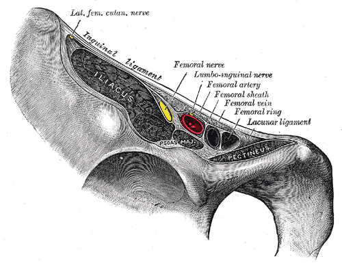
Inguinal ligament/pouparts ligament - is thickening of external oblique aponeurosis and it extends from anterior superior iliac spine to pubic tubercle.
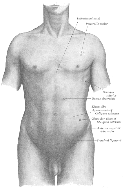
The inguinal ligament (/ˈɪŋɡwɪnəl/[1][2]), also known as Poupart's ligament or groin ligament, is a band running from the pubic tubercle to the anterior superior iliac spine. It forms the base of the inguinal canal through which an indirect inguinal hernia may develop.
The Inguinal ligament
runs from the anterior superior iliac crest of the ilium to the pubic tubercle of the pubic bone. It is formed by the external abdominal oblique aponeurosis and is continuous with the fascia lata of the thigh.
Hope you learned abdomen about the regions, layers and inguinal ligament.in next post we will learn more depth about abdome.stay tuned for it.
References -
Gray' anatomy:the Anatomical Basis of Clinical Practice,41st edition page number 1073
Clinically Oriented Anatomy 7th edition page number 209
Thanks for reading,
With regards

