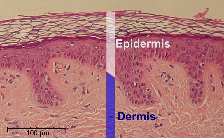
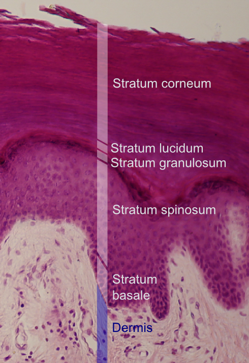
Hello everyone, how are you all? Hope you all are doing well and i am too. Today i would like to focus on skin biopsy. We will get many skin biopsies related to many different disease conditions. One of the most common clinical entity we are going to discuss is "Pemphigus vulgaris".
Pemphigus vulgaris is an autoimmune condition where antibodies are going to be formed against desmosomes. First of all we have to know what are desmosomes. Before that we have to know layers of the skin and what it is made of. Skin has epidermis and dermis. Epidermis has 5 layers and the cells present in epidermis are called keratinocytes. Desmosomes are protein complex present in between keratinocytes and maintain the shape of keratinocytes in polygonal shape.
Desmosomes are many types but here we have to know desmoglein 3 and 1. Desmoglein 3 is more present in lower epidermis compared to desmoglein 1.
So in lower epidermis antibodies are formed against these desmosomes and so these desmosomes will not hold keratinocytes together and there we will see split in the lower epidermis. Patient will present with bullous lesions on skin. So histopathologically what we see i am going to share.
You can see epidermis and the split in the epidermis
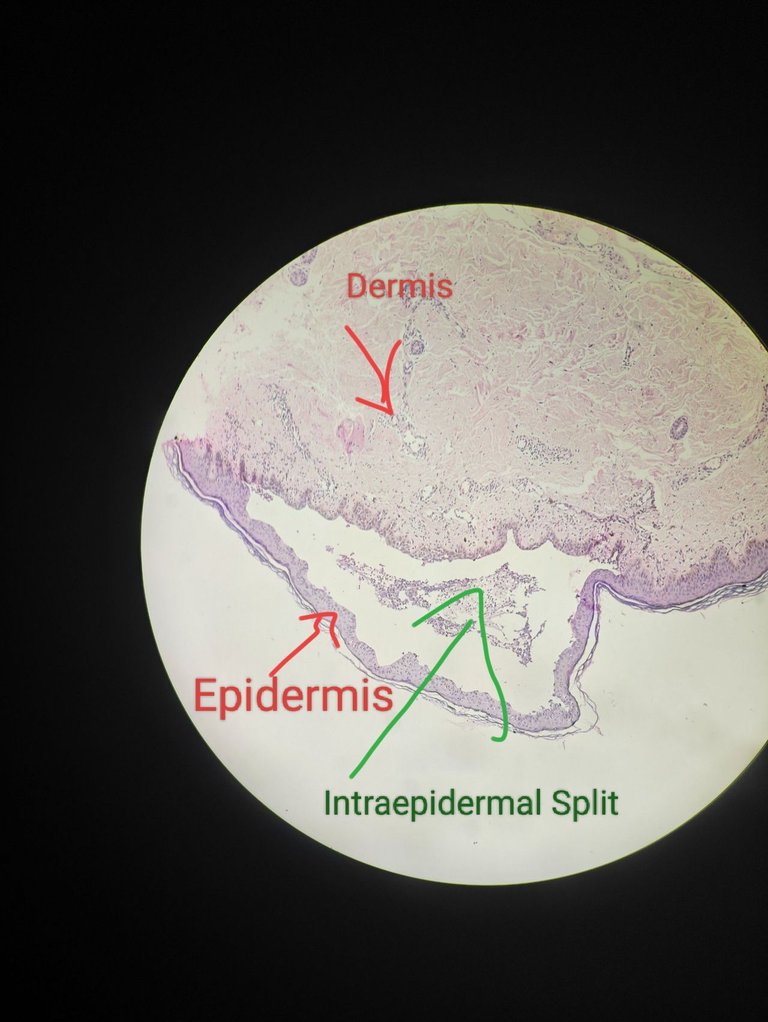
The below images are 100X magnification
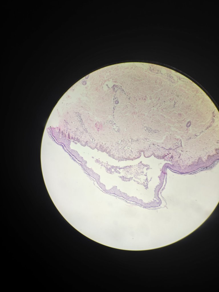
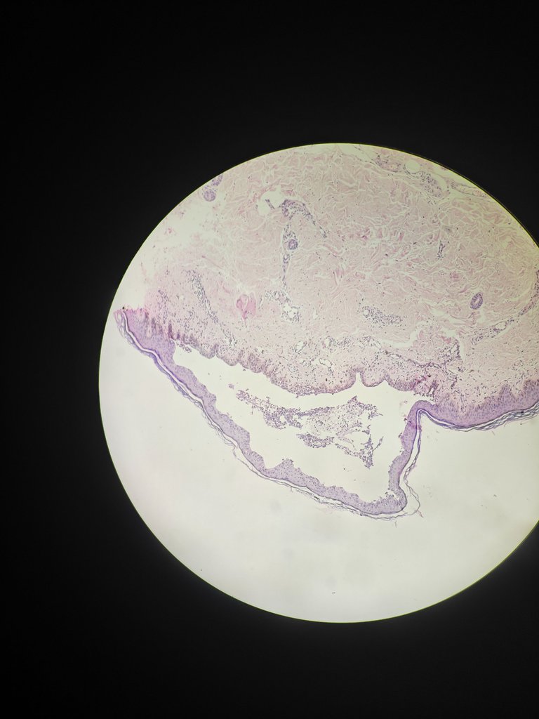
This is 40X magnification
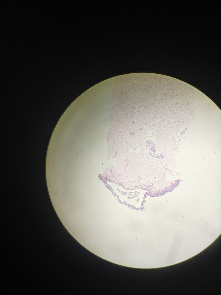
The below image is 400X magnification showing some inflammatory infiltrates collection in the split.
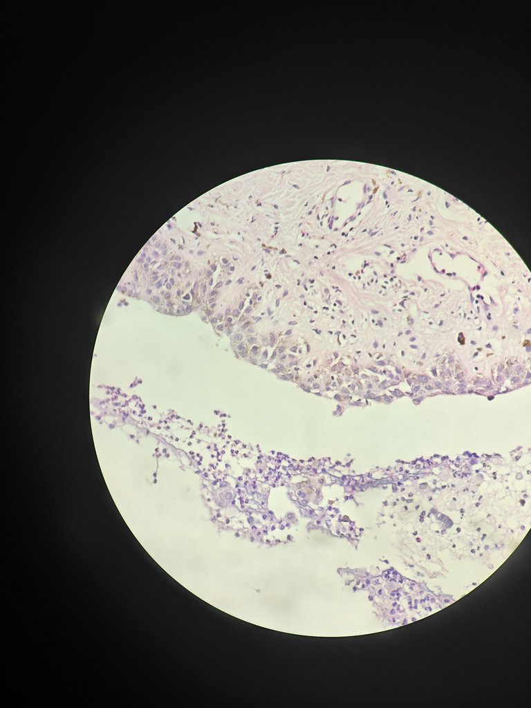
Gold standard is direct immuno fluoroscence but histopathology will give a way to diagnosis
I hope you understood well about how pathology helps in diagnosing skin disorders.
I will come up with another new topic in my next post.
Thanks for reading,
With regards,
References
- Lever's Dermatopathology: Histopathology of the Skin 12th Edition
